Pelleti’s team at the University of Bologna developed a straightforward way to distinguish fly artifacts from real bloodstains using electron microscopy.
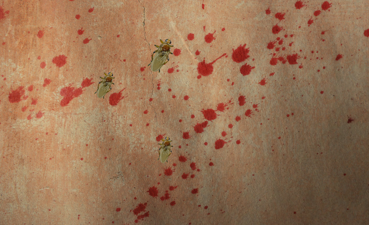
If you have heard of the show Dexter, then you may be acquainted with blood pattern analysis. While the regular blood pattern analyst is not also committing crimes after their day job, they are still quite busy reconstructing the events of a crime by analyzing even the smallest droplets of blood found at a crime scene (Fig. 1).

However, there are many aspects of a crime scene that can potentially hinder blood pattern analysis, including insects. Insects are attracted to a crime scene by the presence of a corpse or blood, both of which can act as their food source. By consuming and interacting with blood stains, insects produce their own stains, known as fly or insect artifacts. These stains can vary in color and shape and are often visually indistinguishable from real bloodstains. This means that a bloodstain analyst may not be able to tell which stains come from a crime and which come from an insect. Some researchers have tried to use biochemical tests that identify blood (like luminol) to differentiate between these types of stains. However, the insect artifacts tend to be heavily comprised of blood, seeing as this is what the insects consumed, and as such can give off misleading results.
A research team from the University of Bologna in Italy wondered if a high-powered microscope that uses electrons to observe samples could be used to visually distinguish between fly artifacts and bloodstains. While these stains might look the same under the naked eye, this powerful type of microscopy, called Scanning Electron Microscopy, can reveal microscopic differences in supposedly flat samples, such as the stains left behind by the interaction of insects with the blood.
Scanning Electron Microscopy, or SEM, works by blasting a beam of concentrated electrons at a sample surface, focused with condenser lenses and deflection coils. The sample will then emit different secondary electrons based on the chemical compounds located on the surface of the sample (Fig. 2). The computer maps these chemical signatures from different regions of the sample to build a microscopic image. SEM photos give both a clear image of tiny objects and information about the sample’s chemical composition, useful for categorizing things by their appearance (the photo itself) and their invisible components (the chemical info).
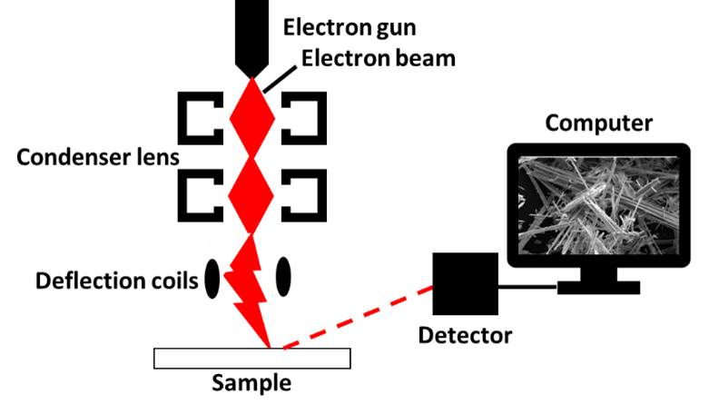
Figure 2. Scanning Electron Microscopy workflow. Credit: Anthophyllite Asbestos Scanning Electron Microscopy (SEM), by Asbestorama on Flickr, edited by Jalissa Thomas.
To test their SEM method, the researchers compared insect artifacts from flies fed human blood to artificially created bloodstains. Two analysts, a forensic pathologist and a biologist with SEM experience, were shown normal camera images, low- and high-magnitude SEM images and asked to characterize the stains. For the camera images, they described basic features of the stains, such as color and shape, and separate fly artifacts into two categories based on these same characteristics. For the SEM images, the experts used a binomial description system for these features: (1) presence/absence of deposits, (2) flat/textured surface, and (3) presence/absence of red blood cells.
The two analysts described the characteristics of stains in the camera and SEM images similarly, showing these types of analyses could be reliable between different scientists. However, many similar features (such as the red-brown color and round shape) were observed across all the stains analyzed. As such, these characteristics would not allow analysts to distinguish between the blood stains and the fly artifacts. However, the three characteristics used for description of SEM images allowed for clear discrimination between bloodstains and fly artifacts (Table 1). The presence of red blood cells was a clear indicator that a stain was pure blood, since fly artifacts do not contain red blood cells. Additionally, both types of fly artifacts have crystal-like deposits, whereas bloodstains did not.

Based on these results, the authors Pelleti et. al. concluded that SEM can accurately identify whether a stain from a crime scene is pure blood or a fly artifact. As industry leaders commercialize more portable SEM instruments that can be transported quickly to crime scenes, this methodology may become a major tool for bloodstain pattern analysts. This can allow experienced and possibly newbie technicians to reliably determine if and to what extent fly artifacts have influenced the bloodstain evidence on-site.
Feature image: “Blood Spatter Texture – FREE” by jumpinjimmyjava, A wallaby fly by Michael Jefferies, edited by Jalissa Thomas.
Citation:
| Title | Scanning electron microscopy in the identification of fly artifacts |
| Authors | Guido Pelletti, Maria Carla Mazzotti, Paolo Fais, Desiree Martini, Laura Ingrà, Alberto Amadasi, Chiara Palazzo, Mirella Falconi, Susi Pelotti |
| Organizations | Department of Medical and Surgical Sciences and Department of Biomedical and Neuromotor Sciences, University of Bologna, Italy |
| Year | 2019 |
| Journal | International Journal of Legal Medicine |
| Link | https://pubmed.ncbi.nlm.nih.gov/31147775/ |

I had no idea that insects can make it hard to tell which stains are from the crime. I feel like a bloodstain analysis service would need to show up quickly. That way they can avoid insects causing problems.
LikeLike