
Trauma analysis is a vital part of any forensic analysis because it gives investigators more context for assessing circumstances of death, working towards a positive identification, or both. Generally, the goals of trauma analysis are to determine the timing of injuries and how they occurred (the “mechanism”). In some scenarios, this process can be relatively straightforward. For example, a single gunshot wound (“high-velocity projectile trauma”) or a single stab wound (“sharp force trauma”) may be easily recognized by the attending pathologist. However, in this case of multiple injuries—particularly those that have occurred over time—the interpretation of trauma can be much more complex. Scientists at the International Manufacturing Center at University of Warwick recently presented three case studies on rib fractures that use Micro-CT, a 3D X-ray imaging technique, to help investigators detect, interpret, and contextualize multiple injuries.
The first goal of skeletal trauma analysis is to determine the mechanism of injuries. Forensic medical investigators analyze and categorize the skeletal trauma as either sharp force trauma, blunt force trauma, or high-velocity projectile trauma. To accomplish this, they assess the pattern that the traumatic injuries leave on bone. For example, a blade (sharp force) will leave a different signature on bone than a baseball bat (blunt force) (Figures 1 and 2).
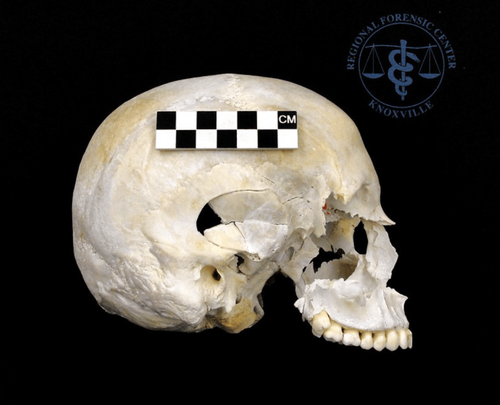
Image available via Future Learn.
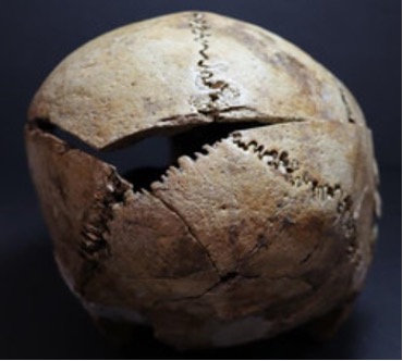
Images available via Future Learn.
The other goal of skeletal trauma analysis is to determine the timing of said injuries. This requires investigators to determine if the injuries occurred antemortem (before death), perimortem (at the time of death), or postmortem (after death).
Antemortem injuries will typically have signs of healing, because the injury occurred during life and the individual’s body was able to respond. In some cases, remnants of antemortem trauma only present as small boney bulges called calluses. However, if an injury is not set properly or disrupted during the healing process, larger remnants are visible. On the other hand, perimortem injuries will have no signs of healing because they occurred at or around the time of death. These injuries are typically what contribute the most to a forensic pathologist’s determination of cause of death.
Unlike with antemortem and perimortem injuries, postmortem injuries are considered taphonomic damage, which can still help investigators understand the circumstances surrounding the death of an individual, but may not actually contribute to understanding the cause of death itself. Collectively, there are many variables that pathologists and anthropologists have to consider simultaneously in trauma analysis. When multiple injuries are introduced, this process only gets more complex.
Drs. Waltraud Baier, Danielle Norman and Mark Williams worked with the West Midlands Police to address one of the most complex and scenarios that forensic investigators encounter—injuries related to child abuse. These can be highly sensitive cases that are not only emotional, but also highly publicized and scrutinized. A focal point of these investigations is often determining if the injuries were accidental. As the authors note, individual injuries are not always characteristic of abuse, so the distribution (i.e. how often and how intense) of injuries is key in separating isolated accidents from reoccurring incidents.
Some studies have shown that ribs will fracture in reaction to CPR-related compressions applied to the front of the ribcage. These injuries would be classified as accidental, rather than intentional abuse. Being able to accurately time when fractures occurred is crucial to identifying abuse. If rib fractures patterns can be linked to multiple, reoccurring incidents, the investigators have a stronger reason to suspect abuse is occurring.
Currently histology, which examines the microstructural organization of tissue, is the gold standard for identifying rib injuries and assessing when they occurred (Figure 3). However, histological analyses can be time consuming and require specific expertise. Other methods for imaging trauma (such as radiographs and ultrasound) are available, but X-ray techniques like micro-CT allows for injuries to be seen in 3D. To showcase the utility of this method to potentially replace or complement histological analysis, Baier and colleagues used micro-CT to identify and categorize skeletal trauma on infant ribcages.

Images from RadiologyKey.
They found micro-CT was able to identify 69% of the fractures identified through histology in addition to 14 fractures not observed through the gold-standard method. This percentage indicates that micro-CT is a promising avenue for fracture detection, but additional refinement is necessary to increase accuracy. However, the authors note that various limiting factors to consider. Most importantly, though micro-CT was able to detect healing fracture calluses, precisely timing when these injuries occurred is currently still dependent on histological analyses. This is particularly true for perimortem fractures that occur before but still close to the time of death, so micro-CT should not be used in isolation just yet. Additionally, they note that some of the additional fractures detected through micro-CT but not observed histologically may represent false positives.
Overall, micro-CT is useful for detecting skeletal fractures and the mechanism of trauma but not a standalone method to determine injury timing. 3D analysis of skeletal trauma can aid histological analysis by directing anthropologists and pathologists to areas that require further analysis. The authors add that the 3D models and videos produced by micro-CT can also help visually demonstrate patterns in injuries occurred to a courtroom, bolstering the understanding of expert testimony. By combining multiple methods, investigators can increase not only the accuracy and precision of their assessments, but also the impact of their testimony. Baier and colleagues’ work provides an important baseline for future studies to build on, allowing forensic scientists to thoroughly pursue allegations of abuse.
| Title | Micro-CT for the examination of paediatric rib injuries: a case series |
| Authors | Waltraud Baier, Danielle G. Norman, and Mark A. Williams |
| Journal | Forensic Science International |
| Year | 2021 |
| Link | https://doi.org/10.1016/j.forsciint.2020.110483 |
| Citation | Baier et al. For. Sci. Int. 2021, 325, 1-6. DOI: 10.1016/j.forsciint.2021. |
